
How to Prove Brain Injury with DTI Imaging After a Normal CT Scan
Traumatic brain injuries alter lives in an instant across New York City. However, standard emergency room imaging often fails to show the true damage. Victims frequently suffer severe cognitive issues while hospital scans report normal results. Consequently, injury claims face aggressive defense challenges. Our Bronx personal injury lawyers have decades of experience helping accident victims build strong medical evidence after serious collisions.
Key Takeaways
- Standard ER CT scans and routine MRIs often miss microscopic diffuse axonal injury in mild traumatic brain injury cases.
- Diffusion Tensor Imaging measures water molecule diffusion along white-matter tracts to reveal microscopic structural damage.
- Lee v. Troge established DTI reliability at trial court level, but Siracusa v. City Ice Pavilion was not verified as establishing this standard.
- In federal DTI admissibility litigation, expert testimony cited that roughly 90 percent of MRI facilities are currently equipped to perform DTI protocols.
You can overcome unhelpful emergency room scans by utilizing advanced neuroimaging. Specifically, Diffusion Tensor Imaging provides clear visual evidence of microscopic brain damage. This step-by-step guide explains how to secure DTI scans and establish liability under New York law.
What You Will Need
- Emergency room discharge records and initial CT scan reports
- A daily log tracking headaches, memory loss, and cognitive struggles
- A referral to a board certified neurologist specializing in traumatic brain injury
- Access to an MRI facility equipped with advanced DTI imaging software
- An experienced legal team to present complex neuroimaging evidence
Step 1: Document Your Initial Symptoms Immediately After the Collision
First, seek immediate medical attention after any crash or severe fall. Emergency room doctors often perform standard CT scans to check for immediate life-threatening bleeding. However, these basic scans usually appear completely normal even when microscopic brain tissue suffers harm.
Meanwhile, you must record every physical and cognitive symptom in detail. Track subtle changes like memory loss, irritability, dizziness, and noise sensitivity. For example, note when light hurts your eyes or when simple tasks feel overwhelming. Detailed personal logs help your doctors connect your symptoms directly to the crash event.
Watch out: Do not assume a normal hospital CT scan means your brain is healthy. Emergency room scans screen for acute surgical emergencies rather than subtle microscopic damage.
Step 2: Recognize the Limitations of Standard Emergency Room Scans
Next, understand why standard hospital equipment misses microscopic brain injuries. Standard emergency room CT scans and routine MRIs frequently miss microscopic diffuse axonal injury in mild traumatic brain injury cases. These standard machines only capture structural anatomical snapshots of larger tissues.
Consequently, defense insurance companies use normal CT scans to argue that victims are exaggerating their injuries. Insurance adjusters regularly point to normal hospital records to deny compensation. Therefore, you must recognize that standard scans are merely the first diagnostic step rather than the final word on your health.
Pro tip: Ask your treating physicians for physical copies and digital files of all emergency room imaging reports. Your specialists will need these early scans to build a clear diagnostic timeline.
Step 3: Consult a Board-Certified Neurologist for Specialized Assessment
Also, schedule a comprehensive evaluation with a board certified neurologist. General practitioners and emergency doctors do not specialize in subtle white matter damage. A specialized neurologist conducts clinical evaluations to measure functional impairments.
For instance, your neurologist will test your working memory, balance, and fine motor skills. In addition, they will evaluate whether your clinical presentation matches diffuse axonal injury. If your symptoms persist beyond two weeks, your neurologist can order advanced neuroimaging protocols.
Pro tip: Request a neurologist who routinely works with advanced neuroimaging protocols. Specialists familiar with court proceedings understand how to properly document brain injuries for trial.
Step 4: Undergo Advanced Diffusion Tensor Imaging Protocols
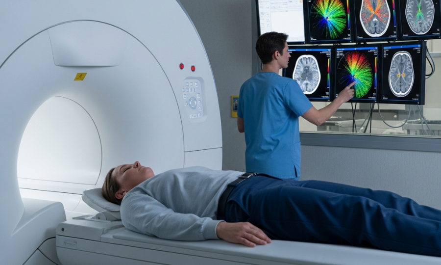
Consequently, secure a referral for Diffusion Tensor Imaging at a qualified facility. Standard MRIs look at tissue structures, whereas Diffusion Tensor Imaging measures water molecule diffusion along white-matter tracts to reveal microscopic damage. When traumatic impacts tear delicate white matter fibers, water molecules diffuse abnormally along those damaged pathways.
Fortunately, access to this technology is widely available today. In federal DTI admissibility litigation, expert testimony cited that roughly 90 percent of MRI facilities are currently equipped to perform DTI imaging protocols. DTI scans create detailed color-coded maps showing exact locations where nerve fibers have been disrupted.
Watch out: Ensure the facility uses dedicated software and proper magnetic resonance sequences. Substandard imaging setups can lead to technical defense objections during trial proceedings.
Step 5: Review Fractional Anisotropy Baseline Values with Your Medical Team
Next, work with your neuroradiologist to review your scan results in detail. Neuroradiologists use fractional anisotropy values as indices of brain fiber integrity, comparing against normative reference values rather than calculating individualized baseline values. Lower anisotropy values in specific white matter tracts demonstrate disrupted cellular pathways resulting from physical trauma.
Defense attorneys frequently attack these numerical baseline calculations in court. However, federal courts consistently reject efforts to throw out this objective medical evidence. The U.S. District Court for the Southern District of New York denied a motion to exclude a neuroradiologist’s DTI testimony, ruling that disputes over fractional anisotropy baseline values go to the weight of the evidence for cross-examination rather than admissibility.
Pro tip: Ensure your neuroradiologist correlates lower numerical values directly with your real world clinical symptoms. Objective visual scans combined with matching physical symptoms create undeniable evidence.
Step 6: Integrate DTI Findings into Your Legal Claim for Trial
In addition, bring your complete imaging files to your legal counsel. Our injury attorneys in the Bronx handle car, construction, and slip-and-fall cases involving complex brain trauma. Your attorney will build a compelling narrative showing how the crash directly caused white matter disruption.
Also, New York trial courts consistently validate DTI technology under state evidentiary standards. In Lee v. Troge, a New York trial court held a Frye hearing and ruled as a matter of law that MRI with DTI is generally accepted in the medical community as a reliable tool for diagnosing brain injuries in individual patients. Similarly, a New York court may consider the admissibility of DTI evidence under the Frye standard to substantiate a plaintiff’s mild traumatic brain injury claim following a motor vehicle accident. Additionally, in Siracusa v. City Ice Pavilion, a New York court quoted prior decisions stating DTI was accepted, but did not independently affirm DTI’s acceptance in the medical community as a reliable diagnostic method for traumatic brain injuries.
Watch out: Defense attorneys often demand raw technical data to delay legal proceedings. However, the New York Supreme Court Appellate Division, First Department, covering Bronx and New York Counties, issued a key discovery decision reversing a trial court order that would have required production of underlying technical data for DTI imaging.
Step 7: Prepare Your Medical Experts to Overcome Defense Challenges
Finally, prepare your treating physicians and expert witnesses for deposition and trial testimony. Defense experts often argue that pre-existing conditions or age caused the white matter changes. Therefore, your experts must explain how the location and pattern of white matter damage match your traumatic impact.
Consequently, clear visual color maps allow Bronx jurors to see microscopic brain damage for themselves. When medical experts explain DTI visual graphics alongside clinical testing, juries gain a complete understanding of your permanent impairment. This step transforms an invisible brain injury into clear objective proof.
Pro tip: Have your medical expert prepare high resolution side-by-side diagnostic visual aids. Showing healthy brain tracts next to your damaged tracts helps non-medical jurors understand the injury instantly.
Next Steps for Bronx Crash Victims
Proving a mild traumatic brain injury requires specialized medical care and decisive legal strategy. Standard hospital CT scans only tell a fraction of the story. By following these steps and securing advanced DTI imaging, you can prove the full extent of your injuries. First, track your symptoms daily. Next, consult a qualified neurologist for advanced imaging options. If you need an experienced Bronx accident lawyer, our firm is ready to fight for you.
Sources
- U.S. District Court for the Southern District of New York, Federal DTI Admissibility and Fractional Anisotropy Rulings
- New York Supreme Court Appellate Division First Department, DTI Imaging Technical Data Discovery Decision
This article was drafted with AI assistance. Please verify all claims and information for accuracy. The content is for informational purposes only and does not constitute professional advice.
Free Consultation
We want to hear about your case! Fill in the form to speak to a Bronx personal injury attorney now!
Follow us for legal tips and case results:
